Understanding Vascular Occlusions
Retinal vascular occlusions occur when blood flow in the retina is blocked, either in a vein or an artery. This can lead to sudden, sometimes permanent vision loss and is a serious condition that requires immediate attention
Categories of Vascular Occlusions:
Artery vs Vein: Occlusions are named based on the type of vessel where the blockage occurs.
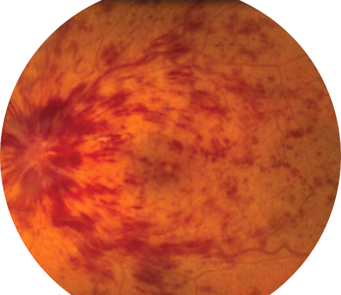
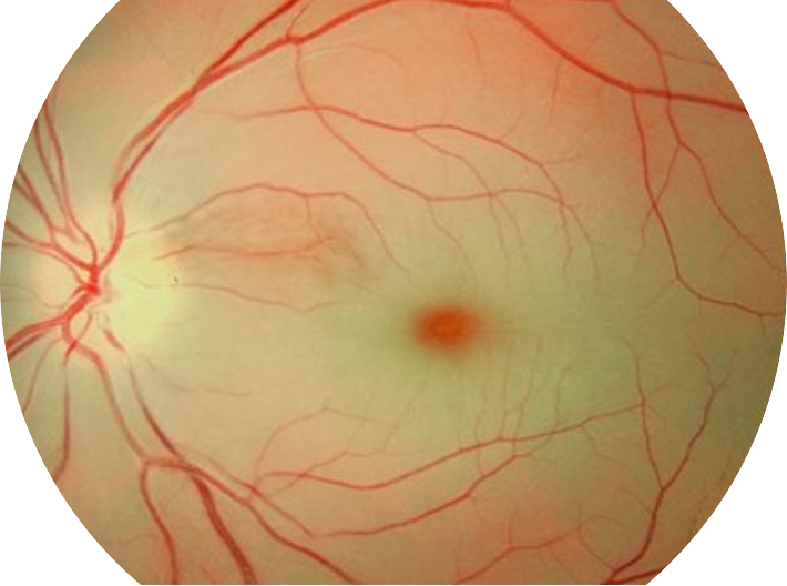
In an Artery Occlusion, blood flow to the retina is cut off and vision loss is often more permanent. In a Vein Occlusion, blood flow out of the retina is interrupted and some vision is more likely to return with treatment
Branched vs Central: In addition to Artery vs Vein, an occlusion is classified by where in the vessel it occurs
In a Central Occlusion, the blockage occurs in the central vessel, and the entire retina is damaged. In a Branched Occlusion, a smaller branch is involved, and only part of the retina is damaged.

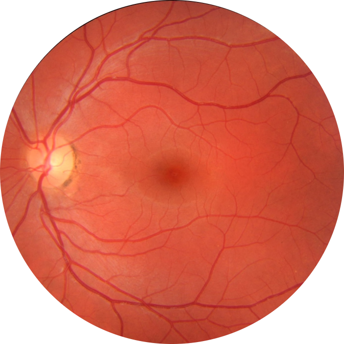
Healthy Eye

Central Vein Occlusion

Central Artery Occlusion
What Causes Vascular Occlusions?
- High Blood Pressure
- Diabetes
- High Cholesterol
- Atherosclerosis (hardening of the arteries)
- Glaucoma
- Blood Clotting Disorders
Symptoms to Watch For:
- Sudden, painless vision loss (partial or complete)
- Blurred or dimmed vision
- Dark spots or blind areas in the field of vision
- Distorted or wavering vision

Normal Vision

Partial Vision Loss from Vascular Occlusion
Diagnosis:
An eye doctor can diagnose retinal vascular occlusion through a comprehensive eye exam, which may include:
- Dilated eye examination
- Optical coherence tomography (OCT)
- Fluorescein angiography (to view blood flow in the retina)
Who’s at Risk?
- Individuals over the age of 50
- Those with a history of cardiovascular disease
- Smokers
- People with high blood pressure, diabetes, or high cholesterol
- Individuals with blood clotting disorders


